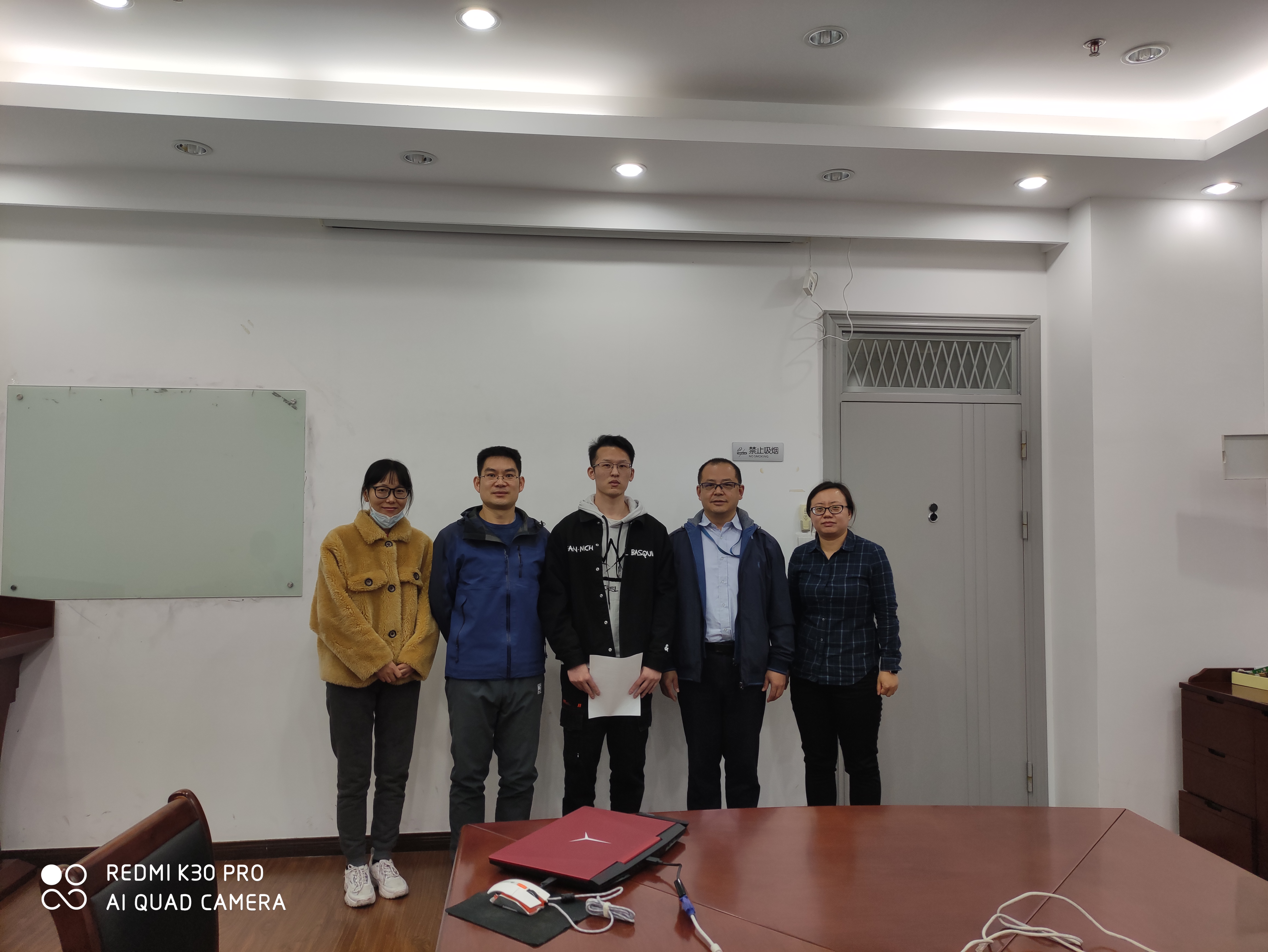
Congratulations to Li Xiaolong on his successful graduation!
Li Xiaolong is a remarkable individual who completed his undergraduate studies at Fujian Engineering College and was admitted to the School of Computer Science at Shanghai University in 2018. Shortly after starting his studies, he joined Professor Han Yuexing’s Image Research Group, focusing on computer vision-related topics. Under Professor Han’s guidance, he chose the topic of medical image segmentation. Over the course of three years, Li Xiaolong worked diligently and utilized various techniques, including deep learning, to achieve automated segmentation of liver and tumor CT images. His contributions have made significant strides in the development of intelligent healthcare. After graduation, Li Xiaolong joined United Imaging Healthcare Co., Ltd., where he continued to work in the field of medical imaging. In addition to his academic pursuits, Li Xiaolong has engaged in extensive extracurricular reading of literature and history. He also has a passion for physical fitness and has formed valuable relationships with mentors and friends. Throughout his three years at Shanghai University, Li Xiaolong has demonstrated tremendous growth. It is hoped that he will maintain his enthusiasm, continuously improve himself, and strive for further breakthroughs in his future endeavors.
Work during graduate studies
In order to assist doctors in making comprehensive assessments and plans for tumor patients, automated segmentation of the liver and tumors in CT images is performed using various deep learning techniques. This provides a data foundation for quantitative and qualitative diagnosis, facilitating clinical treatment. Automated segmentation of the liver and tumors using a combination of deep learning techniques allows doctors to obtain comprehensive evaluations and treatment plans for tumor patients. This automated approach alleviates the workload on doctors and improves the accuracy and efficiency of diagnosis. By utilizing deep learning algorithms, the system can learn and recognize the features of the liver and tumors in CT images, accurately segmenting them. Such segmentation results can be used for quantitative and qualitative assessments, aiding in clinical decision-making.
-
A boundary loss-based 2.5D fully convolutional network (FCN) is proposed to address the limitations of FCN, U-Net, and other deep learning methods in exploring spatial feature information in three-dimensional CT images. This novel approach effectively explores the spatial feature information in CT images while reducing the network parameters and computational resource consumption. The proposed method enhances the accuracy of liver and liver tumor segmentation.
-
To address the characteristics of medical images and the limited capability of common loss functions to optimize network exploration of boundary features, a new boundary loss function is designed. This novel loss function combines the distance, area, and boundary information of image contours, effectively optimizing deep learning networks to explore more image boundary and contour features.
-
To address the issue of encoding-decoding networks ignoring the correlation and dependency among local features, a segmentation framework based on dual-path attention is proposed by integrating 2D, 2.5D, and 3D networks with attention mechanisms. This framework incorporates dual-path self-attention modules, dense network blocks, residual network blocks, and dual-path network blocks. It comprises nine different network structures and is capable of effectively performing automated segmentation of liver and tumor CT images. The integration of these components allows the network to capture and leverage both local and global information, leading to improved segmentation accuracy.

Essay: Research on liver and tumor segmentation using multi-dimensional encoding-decoding networks